
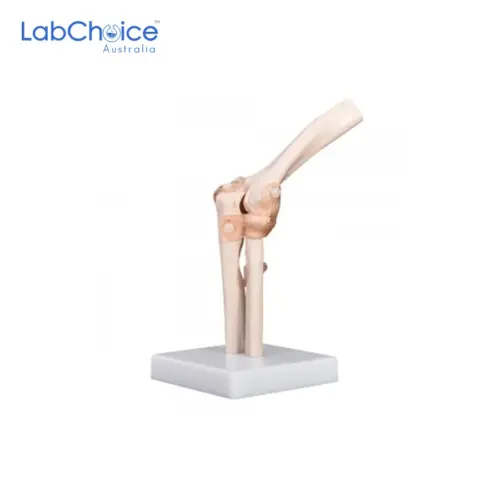




Zoological Frog Dissection Model on Board
AUD 186.90(Ex GST)

Scintillation Vial with White PP Cap, BORO 3.3 Glass
From AUD 200.00 (Ex GST)
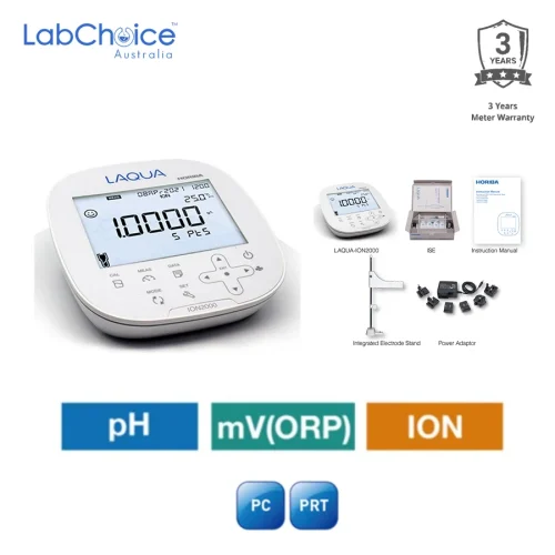
pH Ion Meter Kit with Chloride Testing, pH Range -2.000 to 20.000 pH
AUD 2,495.00(Ex GST)
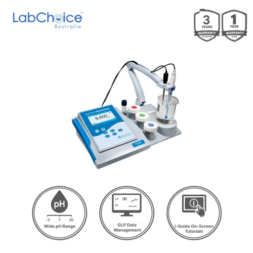
pH Meter Kit PH9500 Research-Grade with TestBench, -2.000 to 20.000 pH
AUD 1,510.00(Ex GST)

Enclosed Low Power Light Sources, 4 mm Connections, 2.5 V MES
From AUD 27.50 (Ex GST)
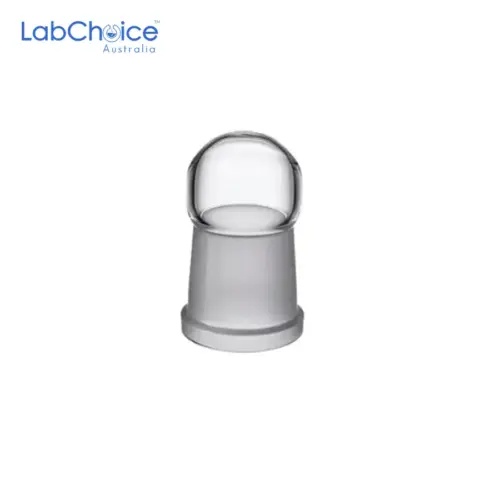

Caps for Schlenk Flasks BORO 3.3 Glass
AUD 32.95(Ex GST)
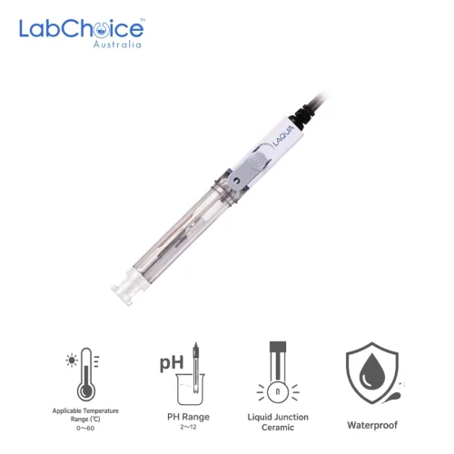
pH Electrode Hydrofluoric Acid Resistant, Pb-Free Plastic Body, 1 m Cable
AUD 295.00(Ex GST)

Dropping Bottle Spare Teats, Red, Pack of 10
AUD 14.90(Ex GST)

PP Caps for Screw Neck Chromatography Vials
From AUD 37.50 (Ex GST)
You May Also Need
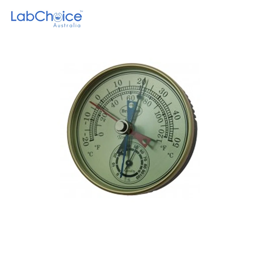
Aneroid Barometer Gilt Dial, for Atmospheric Pressure
AUD 47.50(Ex GST)
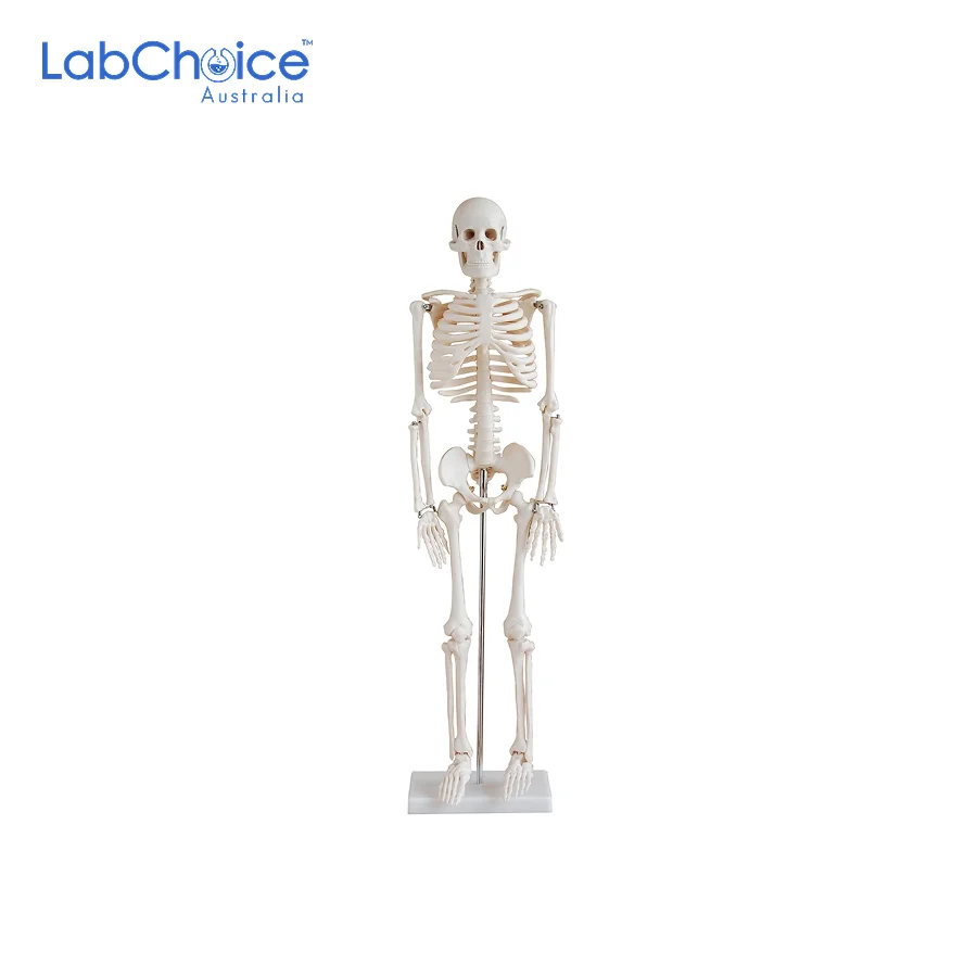
Biological Small Skeleton Model, 850 mm on Stand
AUD 124.50(Ex GST)
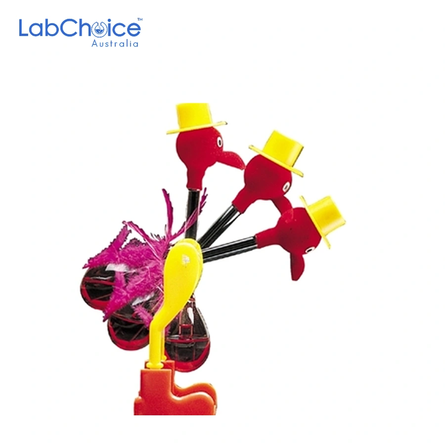
Bird Drinking Heat Thermodynamics Demonstration
AUD 18.90(Ex GST)

Chemistry Periodic Table of Elements with Illustrations
AUD 130.90(Ex GST)
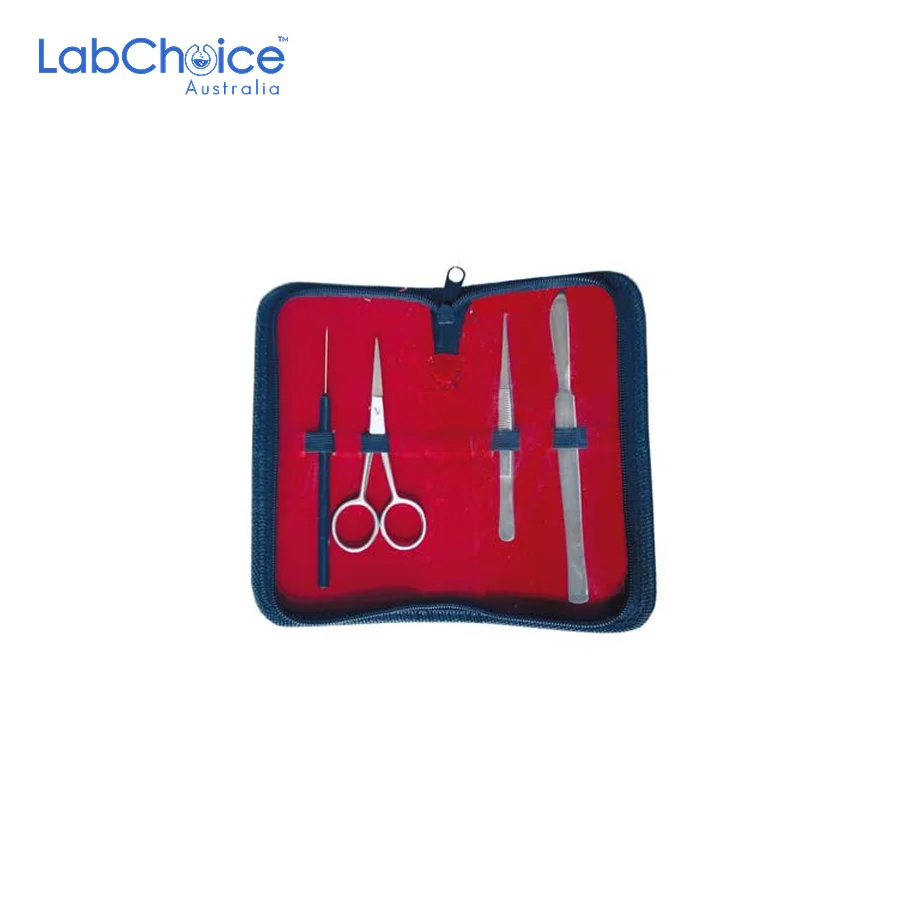
Dissecting Instrument Sets, Walleted Laboratory and Classroom Kits
From AUD 29.90 (Ex GST)
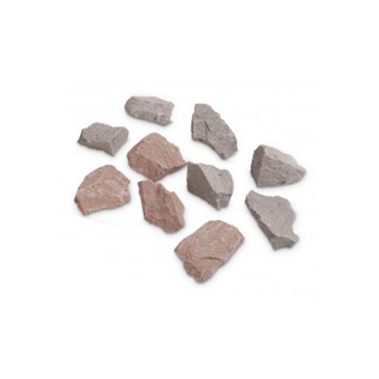
Educational rock specimens For Display
From AUD 27.50 (Ex GST)
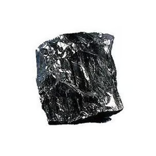
Educational rock specimens for geology
From AUD 24.90 (Ex GST)
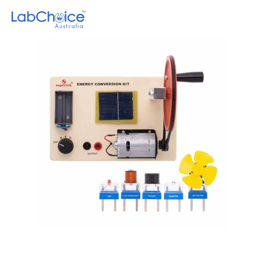
Energy Conversion Kit with Accessories
AUD 174.50(Ex GST)
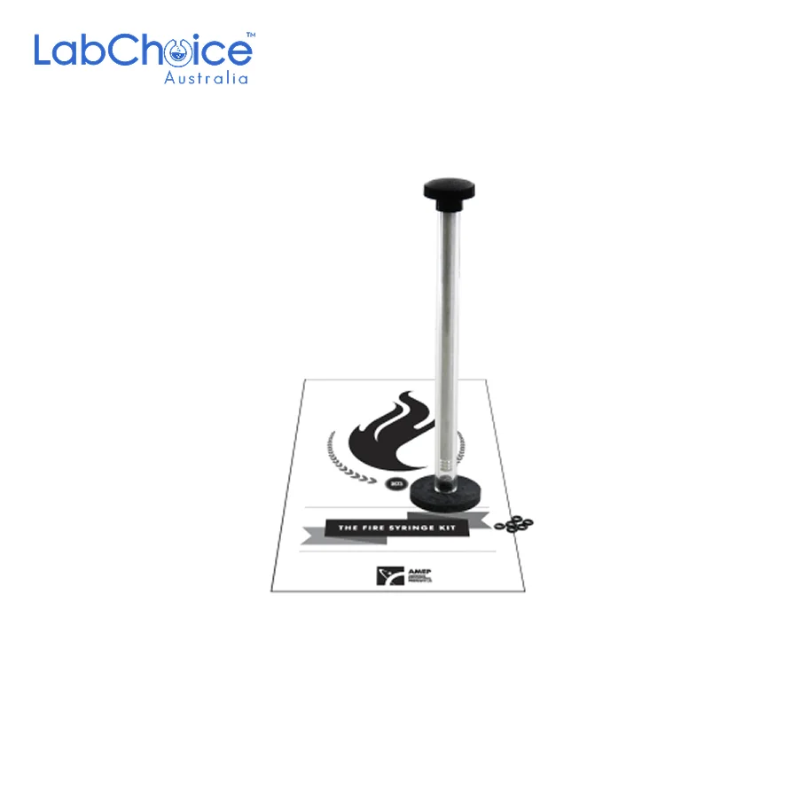
Fire Syringe Kit, Charles’s Law and Compression Demonstration
AUD 74.90(Ex GST)

Individual Mineral Specimens For Display
From AUD 27.50 (Ex GST)
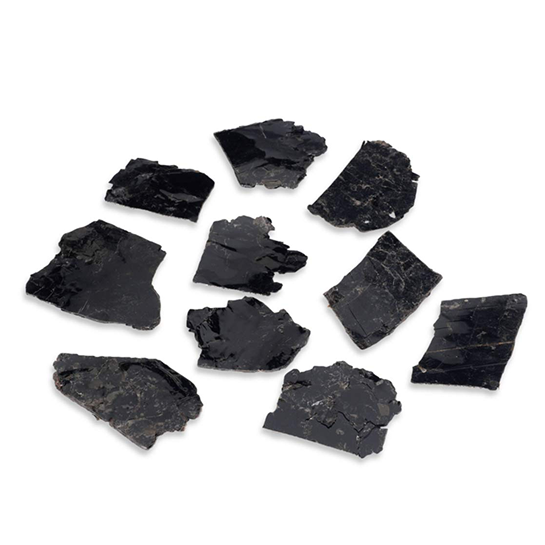
Mineral Specimens for Educational Geology
From AUD 29.90 (Ex GST)
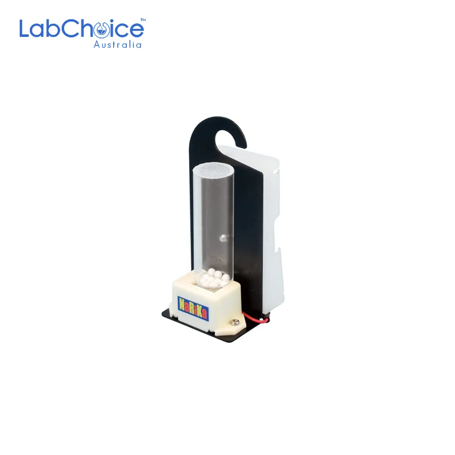
Portable Vacuum Buzzer for Vacuum Experiments
AUD 87.50(Ex GST)

Seismograph Teaching Model, Classroom Seismology Demonstration Set
AUD 83.50(Ex GST)
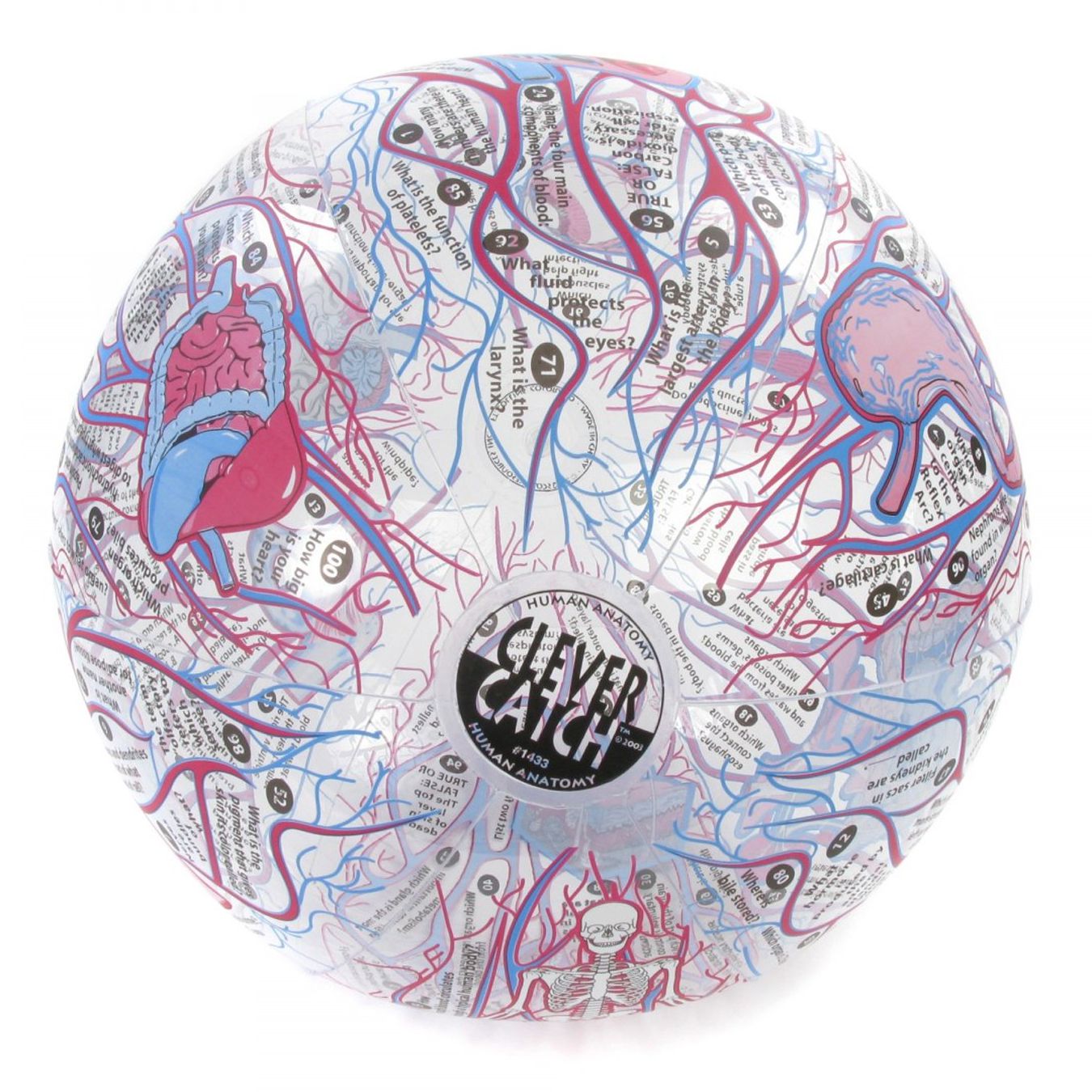
STEM Quiz Balls
From AUD 32.50 (Ex GST)
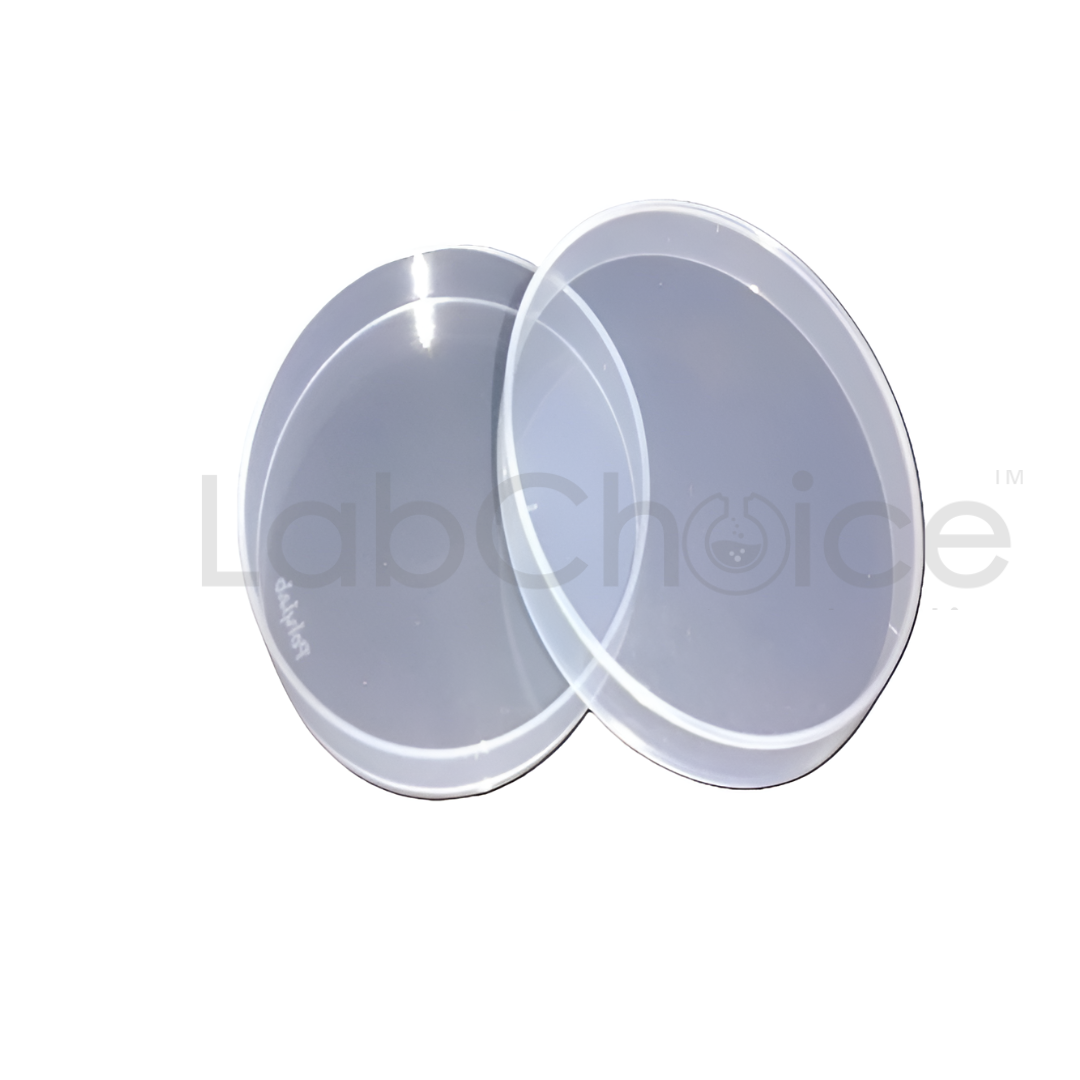
Petri Dishes, Polypropylene, Autoclavable & Reusable, High Quality
From AUD 1.20 (Ex GST)

Stirring Rods, Rounded Ends, Laboratory Grade, Borosilicate Glass (BORO 3.3)
From AUD 0.95 (Ex GST)
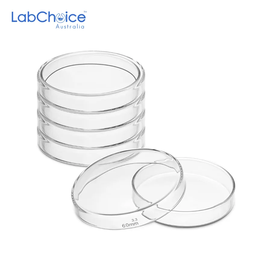

Glass Petri Dishes with Lid, Autoclavable, Borosilicate Glass (BORO 3.3)
From AUD 3.90 (Ex GST)

⭐⭐⭐⭐⭐
We ordered a set of customised BORO 3.3 borosilicate glassware for our teaching labs, and LabChoice Australia exceeded every expectation. From the first consultation to final delivery, their team listened carefully, offered expert advice, and ensured every piece matched our specifications perfectly.
The clarity and precision of the glassware are exceptional, and the quick turnaround made setup effortless. It’s refreshing to work with an Australian supplier who genuinely understands laboratory needs and takes pride in quality craftsmanship. We’ll definitely be returning to LabChoice for future glassware requirements
⭐⭐⭐⭐⭐
Our lab required high-precision BORO 3.3 glassware for analytical testing, and LabChoice Australia delivered outstanding results. The ordering process was simple, communication was clear, and every beaker and flask arrived perfectly calibrated and ready for use.
The quality of the glass is exceptional — resistant to heat, easy to clean, and consistently accurate in measurement. It’s reassuring to know there’s an Australian supplier that combines scientific precision with genuine customer care. I’d recommend LabChoice to any research team looking for dependable labware
⭐⭐⭐⭐⭐
We needed a set of customised borosilicate glassware for a specialised project, and LabChoice Australia delivered exactly what we envisioned. The team worked closely with us from design to production, ensuring every detail met our specifications. The craftsmanship is exceptional, and the turnaround time was impressive. It’s reassuring to know we can rely on them for unique, made-to-order pieces
⭐⭐⭐⭐⭐
LabChoice Australia has completely changed the way we source our lab glassware. The quality is consistently top-notch, and every piece meets the precision we need for reliable results. Their team knows their stuff, answers our questions quickly, and always goes the extra mile to help — they’re more like partners than just a supplier.
⭐⭐⭐⭐⭐
We trust LabChoice Australia because they deliver every time. Their borosilicate glassware is durable, accurate, and perfect for the high-heat work we do. Everything arrives well-packed with clear specs, so there are never surprises. It’s reassuring to deal with a supplier who understands both compliance and the demands of a busy lab